

A detailed test that checks even corneal genes before vision correction
Avellino Genetic Test
You can carry the Avellino corneal dystrophy gene without any symptoms.
Before laser vision correction, a simple sample lets us check in advance.
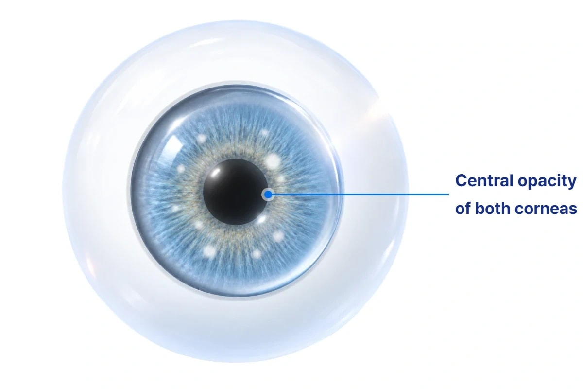
Avellino corneal dystrophy genetic test
Vision correction surgery may involve applying a laser to the cornea or creating a flap.
So before surgery, it is important to look not only at corneal thickness and shape
but also at genetic factors that could affect the cornea.
The Avellino test checks for gene mutations associated with Avellino corneal dystrophy
that may be difficult to detect with standard eye exams alone.
What is Avellino corneal dystrophy?
A hereditary disease in which protein deposits build up in the cornea, gradually reducing vision.
First reported in 1988 in a family from the Avellino region of Italy, it is caused by a gene mutation
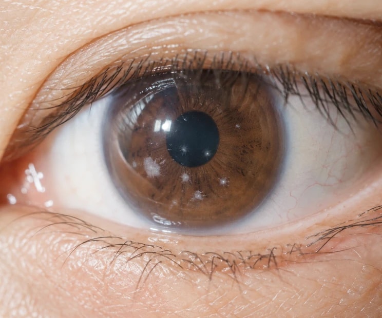
that produces white spot-like opacities in the center of both corneas, which increase with age and reduce vision.
You may not know you carry it until symptoms appear after corneal damage
Hereditary
corneal diseaseWhite spots · opacity
in the central corneaProgressive vision loss
with ageSymptoms appear or worsen
after corneal damageNo symptoms
before onset
Two types of Avellino corneal dystrophy
The age at which symptoms appear and how quickly they progress depend on the gene combination inherited from your parents.
Heterozygous (inherited from one parent)
- Carries the gene on only one of the pair
- White spots begin to appear on the cornea from about age 12
- Spots increase in number and size with age
- Vision declines around age 60–70
- In some people in their 20s and 30s, white spots are not yet visible
Homozygous (inherited from both parents)
- Both genes of the pair are Avellino genes
- Symptoms appear from about age 3
- Progresses earlier and faster than the heterozygous type
Why is an Avellino test needed before vision correction?

You can carry it without symptoms
Most people with this condition go about their lives
without knowing they carry the gene.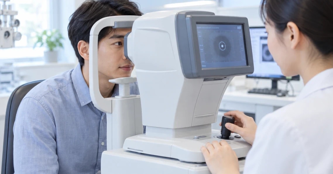
Standard eye exams have limits
Before symptoms appear, the cornea looks much like a normal one, so eye exams such as corneal CT and microscopy
alone have difficulty accurately detecting subtle opacities.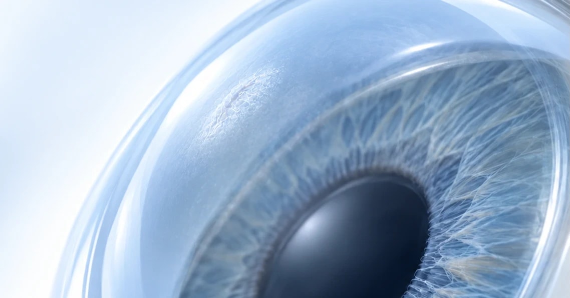
Corneal damage can trigger symptoms
Injury to the cornea can accelerate protein deposits,
so carrier status must be checked before laser vision correction.
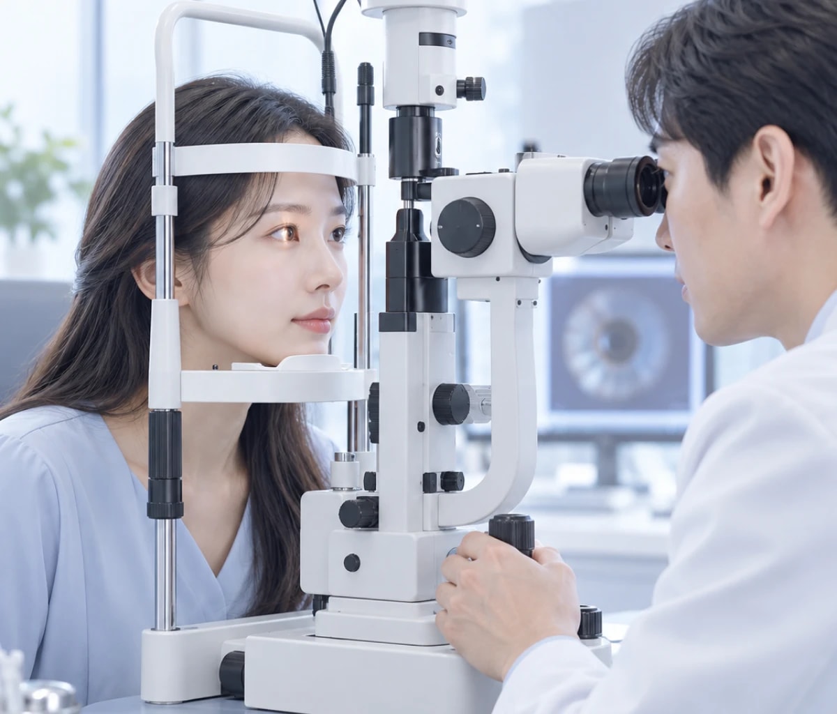
Test Method 01
Slit-Lamp Microscope Exam
A method of diagnosing Avellino corneal dystrophy by directly observing any corneal opacity;
results are available immediately.
- Features
- Immediate results
- Observation only, no sample needed
- Limitations
- Requires diagnosis by an experienced ophthalmologist
- Degree of expression varies with contact lens history, environment and individual differences
- Diagnosis can be subjective, limiting accuracy
Test Method 02
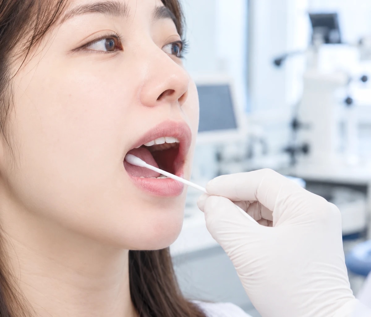
DNA Genetic Test
The genes from collected cells are amplified more than a million times
and analyzed for the Avellino corneal dystrophy gene.
Carrier status can be confirmed objectively, even before symptoms appear.
- Hair Root
5 strands of hair
with roots - Cheek Swab
Simple collection from inside
the mouth with a swab - Blood
Just a tiny amount
of blood drawn by syringe
BGN Eye Clinic Jamsil offers same-day surgery after DNA testing
If this sounds like you, ask us about testing
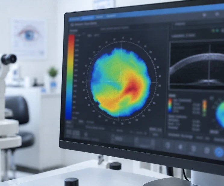
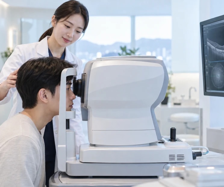
If you are planning vision correction surgery, consider a corneal genetic test as well.
in a previous exam
pre-op examination

such as LASIK or LASEK

with corneal dystrophy
have been seen on your cornea
in a previous exam
pre-op examination
such as LASIK or LASEK
with corneal dystrophy
have been seen on your cornea
in a previous exam
pre-op examination
such as LASIK or LASEK
with corneal dystrophy
have been seen on your cornea
in a previous exam
pre-op examination
such as LASIK or LASEK
with corneal dystrophy
have been seen on your cornea
Frequently Asked Questions
Before symptoms appear it is hard to detect with standard exams, and symptoms can appear after corneal damage, so it should be checked before laser vision correction.
In heterozygous carriers, white spots may not be visible in their 20s and 30s, so you can carry the gene without noticing any symptoms.
Samples are collected simply from hair roots, a cheek swab or blood, so it is easy on you.
The appropriate correction method is decided in consultation with your doctor based on your corneal condition and test results.
BGN Eye Clinic Jamsil offers same-day surgery after DNA testing.